
Page 33 - ESHRE2019
P. 33
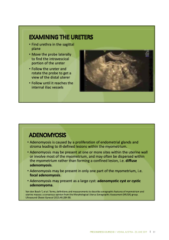
  EXAMINING THE URETERS
• Find urethra in the sagittal plane
• Move the probe laterally to find the intravescical portion of the ureter
• Follow the ureter and rotate the probe to get a view of the distal uterer
• Follow until it reaches the internal iliac vessels
                      ADENOMYOSIS
• Adenomyosis is caused by a proliferation of endometrial glands and stroma leading to ill‐defined lesions within the myometrium.
• Adenomyosis may be present at one or more sites within the uterine wall or involve most of the myometrium, and may often be dispersed within the myometrium rather than forming a confined lesion, i.e. diffuse adenomyosis.
• Adenomyosis may be present in only one part of the myometrium, i.e. focal adenomyosis.
• Adenomyosis may present as a large cyst: adenomyotic cyst or cystic adenomyoma.
Van den Bosch T, et al. Terms, definitions and measurements to describe sonographic features of myometrium and uterine masses: a consensus opinion from the Morphological Uterus Sonographic Assessment (MUSA) group. Ultrasound Obstet Gynecol 2015;46:284‐98.
            28
 PRECONGRESS COURSE 03 I VIENNA, AUSTRIA – 23 JUNE 2019 31
