
Page 88 - ESHRE2019
P. 88
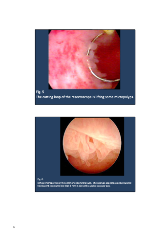
 Fig. 5
The cutting loop of the resectoscope is lifting some micropolyps.
 Fig. 6.
Diffuse micropolyps on the anterior endometrial wall. Micropolyps appears as pedunculated translucent structures less than 1 mm in size with a visible vascular axis.
 86
83
