
Page 102 - ESHRE2019
P. 102
• •
• •
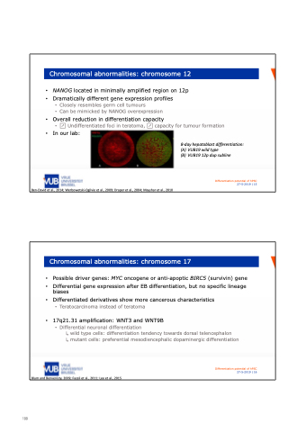
Chromosomal abnormalities: chromosome 12
NANOG located in minimally amplified region on 12p
Dramatically different gene expression profiles
• Closely resembles germ cell tumours
• Can be mimicked by NANOG overexpression
Overall reduction in differentiation capacity
• ↗Undifferentiatedfociinteratoma,↗capacityfortumourformation In our lab:
8‐day hepatoblast differentiation: (A) VUB19 wild type
(B) VUB19 12p dup subline
Differentiation potential of hPSC
27-5-2019 | 15
Ben‐David et al., 2014; Werbowetski‐Ogilvie et al., 2009; Draper et al., 2004; Mayshar et al., 2010
Chromosomal abnormalities: chromosome 17
• Possible driver genes: MYC oncogene or anti-apoptic BIRC5 (survivin) gene
• Differential gene expression after EB differentiation, but no specific lineage
biases
• Differentiated derivatives show more cancerous characteristics
• Teratocarcinoma instead of teratoma
• 17q21.31 amplification: WNT3 and WNT9B • Differential neuronal differentiation
↳ wild type cells: differentiation tendency towards dorsal telencephalon
↳ mutant cells: preferential mesodiencephalic dopaminergic differentiation
Blum and Benvenisty, 2009; Fazeli et al., 2011; Lee et al., 2015
Differentiation potential of hPSC
27-5-2019 | 16
100
97
