
Page 67 - PCC07
P. 67
i
e
i
n
n
P
Po
ol
l
y
y
c
c
y
ys
s
t
ti
i
c
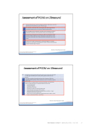
Assessment of PCOM on Ultrasound
CR
Ultrasound should not be used for the diagnosis of PCOS in adolescence, due to the high incidence of multi‐follicular ovaries in this life stage
****
_
CR
The threshold for PCOM should be revised regularly with advancing ultrasound technology and age specific cut off values for PCOM should be defined
****
_
CR
The transvaginal ultrasound approach should be used in the diagnosis of PCOS if acceptable to the patient, with the exception of those not yet sexually active
****
***
_
_
CR
Using ultrasound transducers with a frequency > 8MHz, the threshold for PCOM should be a follicle number per ovary of ≥ 18 and/or an ovarian volume > 10 ml, ensuring no corpora lutea, cysts or dominant follicles are present in one or both ovaries
CPP
In patients with irregular menstrual cycles and hyperandrogenism, an ovarian ultrasound is not necessary for PCOS diagnosis; however ultrasound will identify the complete PCOS phenotype if required
_
_
Th
hi
i
s
sw
w
o
o
r
cO
O
v
va
ar
r
y
yS
S
y
y
n
nd
d
r
ro
om
m
e
e
(
(
p
pr
ro
o
j
je
ec
c
t
tn
n
u
um
mb
be
er
rA
A
e
n
P
P
n
t
P
t
P1
r
re
1
e
f
0
fo
07
o
r
7
rR
8
8
R
4
4
e
es
4
s
44
4
e
e
a
)
).
a
r
.
r
c
c
h
h
E
E
x
x
c
ce
el
ll
le
en
n
c
c
e
Teede et al Human Reproduction 2018
T
r
k
ki
is
s
s
s
u
u
p
pp
po
or
r
t
t
e
e
d
db
by
y
t
th
he
eN
N
H
H
M
M
R
R
C
C
f
fu
u
n
n
d
d
e
ed
dC
C
e
i
e
i
n
n
P
Po
ol
l
y
y
c
c
y
ys
s
t
ti
i
c
cO
O
v
va
ar
r
y
Assessment of PCOM on Ultrasound
CPP
Transabdominal ultrasound should primarily report ovarian volume with a threshold of > 10ml, given the difficulty of accurately assessing follicle number with this approach
_
_
CPP
If using lower resolution ultrasound transducers with a frequency < 8MHz, the threshold for PCOM should be a follicle number per ovary of ≥ 12 and/or an ovarian volume ≥ 10ml
_
_
CPP
Clear protocols are recommended for reporting antral follicle count and ovarian volume on ultrasound. Minimum reporting standards should include:
Last menstrual period
Transducer frequency
Approach/route assessed
Total follicle number per ovary measuring 2‐9mm
Three dimensions of each ovary and the volume
Reporting of endometrial thickness and appearance is preferred – 3 layer
endometrial assessment may be useful
Other ovarian and uterine pathology including ovarian cysts as well as corpus
luteum, and dominant follicles > equal 10mm
_
_
yS
S
y
y
n
nd
d
r
ro
om
m
e
e
(
(
p
pr
ro
o
j
je
ec
c
t
tn
n
u
um
mb
be
er
rA
A
P
P
P
P1
re
1
e
f
0
fo
07
o
r
7
rR
8
8
R
4
4
e
es
4
s
44
4
e
e
a
)
).
a
r
.
r
c
c
h
h
E
E
x
x
c
ce
el
ll
le
en
n
c
c
e
Teede et al Human Reproduction 2018
T
Th
hi
i
s
sw
w
o
o
r
r
k
ki
is
s
s
s
u
u
p
pp
po
or
r
t
t
e
e
d
db
by
y
t
th
he
eN
N
H
H
M
M
R
R
C
C
f
fu
u
n
n
d
d
e
ed
dC
C
e
e
n
n
t
t
r
Page 60 of 196
PRECONGRESS COURSE 07 I BARCELONA, SPAIN – 1 JULY 2018 67
